Remap GMI

Small steps toward comfortable movement.

Staging build · Test accounts and test patients only.

Remap GMI

GRADED MOTOR IMAGERY

Welcome back.

Sign in to your practice, or enter the code from your therapist.

GMI Clinic · Account

Patient sign in

Use the email and password you created when joining.

Have a personal code?

Use the personal code assigned by your therapist to set up your account.

GMI Clinic · Enrollment

Join your clinic

Enter the patient code from your therapist, then create your login. If you opened the link they sent, the code is already filled in.

20 letters and numbers, for example ABCDE-FGHJK-MNPQR-STVWX. Dashes and spaces are optional. Each code works once, for you only.

Your account uses a code in the clinic dashboard. Do not enter your name or medical history here.

Account setup needed

This account is not linked to a clinic. Enter the patient code from your therapist, or ask the clinic administrator to complete staff setup.

Clinic · Clinician

Patients

Active patients—in this view
Practiced this week—last 7 days
Avg accuracy—left/right, last 7 days
Avg response time—left/right, last 7 days
Show

GMI Clinic · Staff

Join your clinic team

Your clinic administrator sent you a personal staff invite link. Create your therapist login below.

Filled in from your invite link: 20 letters and numbers. Each invite works once.

Remap GMI · Platform owner

Remap GMI · Platform owner

Owner dashboard

Create clinics, manage seats, and open the shared photo and imagery-audio tools.

Create a clinic

3 to 40 lower-case letters, numbers and dashes. Filled in from the name; it cannot be changed later.

Usually 3 to 15 (1 to 50 allowed). Every active therapist uses one seat. Clinic admins do not use a seat unless they also treat patients, so the first admin can always join.

Patient review

Patient

Current plan

Left/right recognition
—
Imagery
—

Recent practice

Loading…

Key measures

Loading…

Document

Copy-and-paste notes for the EMR. They use the dates and settings chosen on the Results tab (by default the last 30 days).

My Results

Left/right recognition

Imagined movement

Welcome back.

Your practice plan for today.

Your practice

Left/right recognition · Level 1

Clinic starting plan

Imagine movement

Imagined movement

Today’s practice

Practice tips

Other practice

Left/right recognition for another body part

Therapist ·

Training photo library

Platform owner only. These photos are shared by every clinic. Give each photo a body part, a side and a level.

Levels
Library coverage: what each level still needs

Patients can practise a level once it has at least 4 labelled photos for that body part, including at least 1 left and 1 right. More photos mean less repetition (sessions use 10–50 trials). Unsaved label changes are not counted.

Add photos

Choose one or many photos (JPEG, PNG or WebP, up to 10 MB each). Set the body part, side and level for each photo, then upload. Labels found in a file name (for example hand-left-L3.jpg) are filled in for you to check.

Choose photos to upload, or finish labeling photos below.

Optimise existing photos

Makes a smaller display copy (up to 1800 pixels on the longest side) of each saved photo that does not have one yet, so practice photos load faster. The original photos are kept exactly as they are: nothing is replaced or deleted. You can stop at any time and run it again later; it carries on with the photos still missing a copy. New uploads get their copy straight away.

Photos to label

Photos uploaded without a full set of labels, and photos you chose to relabel, appear here. Fully labelled photos are in the saved library.

All changes saved

Therapist dashboard · Photo library

Saved photos

Choose a body part, then difficulty level, then left or right to see the saved photos.

1. Body part

Removing a staging Firebase photo deletes it from the practice library and Firebase photo storage. Older hosted photos are only removed from the practice library.

Shared library · All clinics

Imagery audio recordings

Session setup

Keep practice comfortable. Pause or end the session if symptoms rise; there is no need to finish every image.
Trial 1 of 20
Preparing image...

Keyboard: press ← or L for left, → or R for right.

Choose accurately at a comfortable pace. Stop if symptoms rise.

Your results

Share lasting changes with your therapist. Responses are not monitored in real time.

Understand · Practice · Progress

What is Graded Motor Imagery?

A step-by-step approach to practicing movement, starting in your mind.

Graded Motor Imagery, or GMI, is a rehabilitation approach for persistent pain and movement difficulties. Think of it as a step-by-step exercise program for the brain: you begin by recognizing body parts, then imagine movement, and later explore mirror feedback with your therapist.

It draws on neuroplasticity—the brain’s ability to adapt through experience and practice. “Graded” means starting with a manageable task and gradually increasing its complexity, according to your response. The aim is to build confidence and comfortable movement without repeatedly provoking symptoms.

What is its purpose?

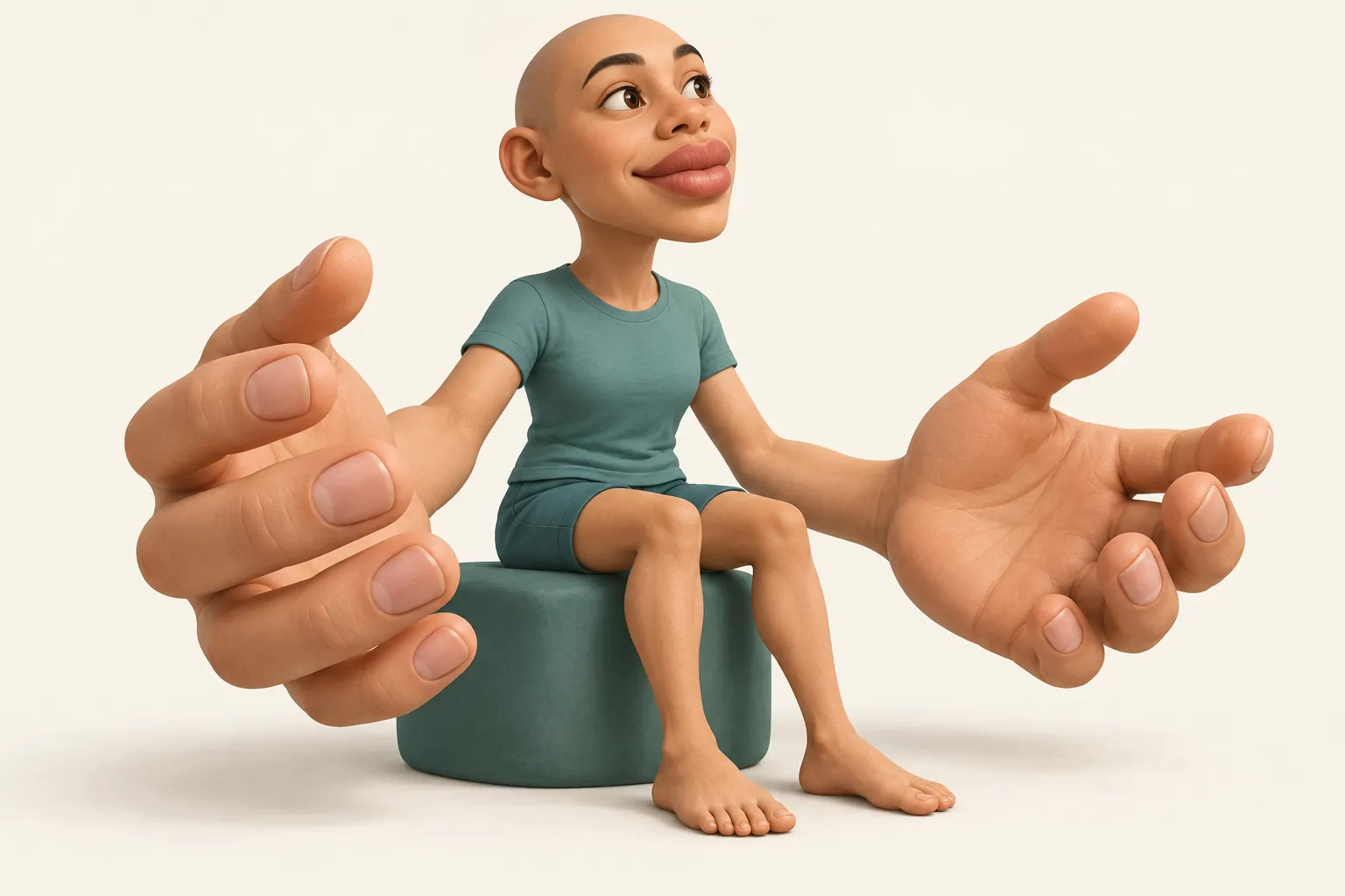
The goal is to reduce pain and help you use your body more confidently. Persistent pain can involve a more protective nervous system and changes in how a body part feels, is recognized, or is imagined moving. People sometimes describe this as a “smudged body map.” That is a useful analogy, not a diagnosis or a sign that the pain is imaginary. [8]

The somatosensory cortex helps process touch and body position. It is one part of the brain’s body-mapping system. GMI aims to help this system and connected movement networks work more normally. Repeated, manageable practice may help movement feel more familiar and less threatening, potentially contributing to pain relief. [12]

Stylized sensory homunculus with enlarged hands and lips and a smaller torso, illustrating unequal sensory representation in the brain.
Your body, represented by sensationThis is a simplified sensory homunculus. Its oversized hands and lips illustrate how some body parts occupy more space in the brain’s touch map than their physical size would suggest.AI-generated conceptual illustration; proportions are illustrative, not measured. This shows a general principle of sensory representation—not a map of pain or damage.

After amputation, the brain can still represent the missing limb, even though its sensory input has changed. Research shows that this representation can remain stable. GMI does not require us to assume that everyone’s map is damaged or that pain relief depends on “repairing” it. [9]

The three stages

How does it work?

Your therapist can tailor the sequence and pace to your needs. These stages gradually introduce recognition, mental rehearsal, and visual feedback.

Left/right recognition

Recognize · Left/right recognition

Look at a picture of a hand, foot, or shoulder and decide whether it is left or right. You do this without moving the affected body part. The task engages body-recognition and movement-planning networks and gives them a manageable starting exercise.

Some people with persistent pain find these judgments slower or harder. Practice prepares you for imagined movement; a score alone does not tell us whether your body map is normal. [8]

Explicit motor imagery

Imagine · Rehearse movement

Imagine moving your affected body part while the actual limb rests. For example, picture opening your hand, turning your palm, or feeling your fingers curl around a cup. Try to imagine the feel of a familiar, comfortable movement—not just watch yourself from a distance.

Imagined and actual movement activate overlapping brain networks. Imagining how a movement feels can also engage somatosensory areas. The activation patterns are similar in some respects, but are not identical. [10]

Mirror therapy

Reflect · Explore visual feedback

A mirror hides the affected or missing limb and reflects the other limb. Watching its reflection can create the impression that the affected side is moving comfortably. This provides a different combination of visual and movement information for the brain to process.

Your therapist helps you position the mirror and choose suitable movements. Mirror therapy may support pain relief or movement recovery, but the response varies. Guided mirror practice in this app is still being developed. [1]

A familiar example: athletes. Athletes mentally rehearse the timing, position, and feel of a basketball shot or volleyball serve before performing it. This rehearsal uses some of the same brain networks as physical movement. GMI applies a similar idea to everyday actions: practicing movement in your mind before building toward physical activity. Athletic studies illustrate mental rehearsal; they do not establish pain-treatment benefits. [11]

Patience, repeatable practice, and regular conversations with your therapist help you find an approach that fits. Progress is about everyday comfort and function, not completing stages on a deadline.

Go deeper · Optional readingA thorough review of research on Graded Motor Imagery and mirror therapyExplore how it may work, findings by condition, and what remains uncertain.

Neurobiological mechanisms and brain-imaging evidence

GMI targets how sensory and movement networks process a body part. Brain-imaging studies have found differences in somatosensory and motor processing in some people with persistent pain. However, findings vary across conditions and people; a single “smudged map” explanation does not account for everyone’s pain. [8][9]

Laterality exercises engage recognition and movement planning. Explicit imagery rehearses a movement without performing it. Mirror therapy provides visual feedback that may help the brain integrate what it sees with movement intention and body sensation. These are plausible, overlapping mechanisms; mirror neurons alone do not explain all of GMI’s effects. [10]

A small randomized crossover study in CRPS found that six weeks of GMI reduced movement pain alongside changes in somatosensory activity and hand representation. This supports the idea that sensory and movement processing can change during rehabilitation. It does not establish that cortical “normalization” caused the pain relief, or that everyone needs the same brain changes to improve. [12]

Complex regional pain syndrome (CRPS)

GMI is a rehabilitation option for CRPS. Small trials and reviews report improvements in pain and disability, sometimes persisting at follow-up. Donati and colleagues’ 2024 review reported benefits from GMI and mirror therapy, while noting small samples and variable study designs. [13]

Confidence in the size of benefit differs between reviews. A 2024 motor-imagery review was more favorable, while the broader 2022 Cochrane review rated the physiotherapy evidence as very uncertain. Swelling has improved in some studies, but GMI is not an established stand-alone edema treatment. We cannot promise a particular reduction in pain, swelling, or disability. [2][3]

Stroke rehabilitation

Mirror therapy has a broader research base for stroke-related movement difficulties than the complete three-stage GMI program. A Cochrane review found moderate-quality evidence of improved motor function and daily activities when mirror therapy was compared with other interventions. It can supplement conventional rehabilitation; findings for mirror therapy should not automatically be attributed to GMI as a whole. [15]

Studies include people at different stages of recovery. Vision, attention, comprehension, fatigue, and movement ability affect how practice should be adapted. Evidence does not establish one best schedule for every stroke survivor.

Phantom limb pain

GMI and mirror therapy may reduce phantom limb pain for some people. A 2023 systematic review found promising effects, but the certainty was very low. Mirror therapy did not show a clear advantage over a covered-mirror control in the pooled comparison, and imagined movement alone did not show a clear benefit. These findings leave uncertainty about which components help most and why. [14]

Some individual trials have reported benefit from repeated mirror practice. The full program remains an option to discuss, rather than a guaranteed treatment or proof that the missing limb’s map must be restored. [16][9]

Chronic low back pain

Research on GMI alone is limited. A larger trial of graded sensorimotor retraining reported improved pain, but its program combined education, sensory training, imagery, and functional movement practice. We cannot assume the same result from laterality or imagery alone. [17]

Chronic shoulder pain and frozen shoulder

Early studies suggest possible benefits when GMI is added to rehabilitation. A small frozen-shoulder pilot trial examined GMI alongside conventional physiotherapy. Such findings are encouraging, but small studies cannot establish reliable long-term benefit or the best protocol for all shoulder conditions. [18]

Knee and hip osteoarthritis

Evidence is developing. A knee-osteoarthritis trial studied GMI alongside exercises and reported benefits in selected outcomes compared with another treatment plus exercises. That does not establish GMI as a replacement for physical rehabilitation, nor does a knee study establish the same benefit for hip arthritis or recovery after joint replacement. [19]

What the evidence means for your practice

GMI and mirror therapy are useful options to discuss within rehabilitation. Benefits and evidence certainty differ by condition. The complete program, its individual components, and this app are not interchangeable: these specific app exercises have not been clinically validated. The aim is a manageable path toward better function, with your response guiding adjustments.


Make practice part of your day

Recommended treatment protocols and adherence

Build a plan with your therapist that you can repeat consistently. Research uses a wide range of schedules and has not established one ideal dose—or a safe daily repetition limit—for everyone. Studied schedules are examples, not targets you must meet. [4]

Start manageable

A suggested laterality starting plan is 4–6 short sessions each day, 2–5 minutes each, with around 30–50 images. Your therapist can adjust the plan. Image count and time are separate guides; more is not automatically better.

Progress by response

Consider symptoms during practice and later that day. Increase one element at a time with your therapist’s guidance. A laterality score alone does not decide readiness.

Build a routine

Link practice to a regular break or daily habit. Start in a quiet setting. As agreed with your therapist, vary familiar safe settings and naturally occurring moods once comfortable. Do not deliberately create distress or practice while driving.

Track what matters

Note what you practiced, how long, and how you felt afterward. Review comfort and everyday function with your therapist—not just speed or accuracy.

Left/right recognition: a starting example

About 30–50 images per session, 4–6 short sessions daily. A suggested starting plan your therapist can adjust, not a standard. Image count and session time are separate measures.

Imagery: a starting example

1–2 minutes, once or twice daily. You might begin with 3–5 imagined movement cycles, with pauses, or less if needed.

These are adaptable app starting examples, not research-validated prescriptions or safety limits. Follow your clinician’s plan. There is no need to finish a target when symptoms rise.

If symptoms change

Even imagined movement can sometimes increase pain or swelling. Pause or stop if pain builds, or if dizziness, confusion, distress, or a sense of disconnection develops. Let symptoms settle and discuss a shorter task, simpler movement, or earlier tolerated stage with your therapist.

Contact your therapist before progressing if practice causes repeated flares or lasting worsening. New or unusual symptoms, marked swelling, or new weakness need clinical assessment. Follow surgical, fracture, and movement precautions throughout. [6][7][5]

Explore guidance for your condition

Open only the sections relevant to you. These describe research examples and clinical considerations, not an individual prescription.

Complex regional pain syndrome (CRPS)A graded sequence, adapted to your response

Research example: a six-week program with two weeks each of laterality, explicit imagery, and mirror therapy. Some original trials used frequent short practice periods, including every waking hour. This intensive schedule is not a starting requirement. [1] [4]

Discuss which stage is manageable and how to adapt daily practice. Do not advance just because two weeks have passed. Your therapist may reduce exposure or return to an earlier tolerated stage if symptoms repeatedly increase.

Stroke-related movement difficultiesMirror therapy alongside rehabilitation

Research example: the stroke mirror-therapy studies averaged about 30 minutes per session, five days weekly, over four weeks, with substantial variation. This describes mirror therapy, not a proven dose for all three GMI stages. [15]

A stroke rehabilitation clinician should adapt practice to vision, attention, comprehension, fatigue, and movement ability. Shorter supported sessions may be appropriate; the study average is not a required starting dose.

Phantom limb painStart with a comfortable sense of the limb

Research example: one small upper-limb mirror-therapy trial used 15 minutes daily, five days weekly, for four weeks. This is a studied schedule, not a guaranteed effective or safe dose for everyone. [16]

Work with your therapist on whether to begin with laterality, imagery, or mirror feedback. Start with simple, familiar movements and adjust if pain or emotional distress increases. Imagery means mentally rehearsing movement; it does not require forceful phantom movement.

Chronic low back painA broader rehabilitation plan

No established GMI-only schedule. Research on broader sensorimotor retraining includes multiple treatments, so its results cannot be assigned to imagery alone. [17]

Your therapist can decide whether imagery fits alongside education and physical rehabilitation. This app currently has hand, shoulder, and foot practice; it does not provide a back-specific training program.

Shoulder pain and frozen shoulderAn optional addition to shoulder rehabilitation

Research example: a small frozen-shoulder pilot trial added GMI to conventional physiotherapy three times weekly for three weeks. It does not establish an optimal home-practice dose. [18]

Begin with an imagined action that feels manageable, such as a supported reach. Your therapist should adapt the plan to your diagnosis and movement restrictions. Imagery supplements the shoulder rehabilitation plan rather than replacing it.

Knee or hip osteoarthritisIndividualize imagery alongside exercise

Research example: a knee-OA trial studied an eight-week program with exercises in both treatment groups. This does not establish a universal daily dose or the same effect for hip OA. [19]

Your therapist can advise whether movement imagery fits your exercise plan. This app does not yet include dedicated knee or hip exercises. After joint replacement, follow your surgical precautions and prescribed rehabilitation.

Need help following the plan? Bring your notes to your therapist and choose a smaller, repeatable routine together. A missed session is a reason to resume your usual plan, not to double the next session.

Evidence behind the guideReferences & further readingOpen the research papers and patient guidance.

References are provided for transparency. Trials, reviews, and patient guidance answer different questions; a published schedule is not an individualized prescription.

  1. Moseley (2006). Graded motor imagery for pathologic pain: a randomized controlled trial. Full GMI program; CRPS and phantom limb pain.
  2. Ríos-León et al. (2024). Effectiveness of motor imagery in complex regional pain syndrome. Systematic review and meta-analysis.
  3. Smart et al. (2022). Physiotherapy for pain and disability in adults with CRPS. Cochrane review.
  4. Williams et al. (2026). Prescription of graded motor imagery for people with chronic pain. Scoping review of dosing and delivery.
  5. European Pain Federation. Explicit motor imagery. Clinical practice guidance.
  6. Moseley et al. (2008). Thinking about movement hurts. Study of symptom responses to imagery.
  7. Northern Care Alliance NHS. Hand therapy: graded motor imagery. Patient guidance on practice and symptom changes.
  8. Halicka et al. (2020). Neuropsychological changes in CRPS. Body perception and laterality recognition.
  9. Stable cortical body maps before and after arm amputation (2025). Longitudinal brain-imaging research.
  10. Target-oriented motor imagery for grasping action (2019). Functional MRI study.
  11. Neural efficiency and acquired motor skills: an fMRI study of expert athletes (2019). Imagined basketball and volleyball skills.
  12. Strauss et al. (2021). Graded motor imagery modifies movement pain, cortical excitability and sensorimotor function in CRPS. Small randomized crossover study linking clinical improvement with sensory and motor brain changes.
  13. Donati et al. (2024). Breaking the cycle of pain: the role of graded motor imagery and mirror therapy in CRPS. Systematic review; benefits reported, with small and varied studies.
  14. Limakatso et al. (2023). The efficacy of graded motor imagery and its components on phantom limb pain and disability. Systematic review and meta-analysis; low-certainty findings.
  15. Thieme et al. (2018). Mirror therapy for improving motor function after stroke. Cochrane review; mirror therapy evidence and treatment schedules.
  16. Finn et al. (2017). A randomized controlled trial of mirror therapy for upper extremity phantom limb pain in male amputees. Small trial; 15 minutes, five days weekly, for four weeks.
  17. Bagg et al. (2022). Effect of graded sensorimotor retraining on pain intensity in chronic low back pain. Randomized trial of the broader RESOLVE program; not GMI alone.
  18. Effectiveness of graded motor imagery in subjects with frozen shoulder (2022). Pilot randomized trial; GMI added to conventional physiotherapy.
  19. Kızkın et al. (2025). The effect of graded motor imagery on pain and function in knee osteoarthritis. Comparative randomized trial; both groups also performed exercises.

Content reviewed September 2026.

Ready for your next manageable step?

A plan that fits you

My practice range

Enter the range you discussed with your therapist, or use these examples as a starting point for that discussion. You can do extra laterality sessions when comfortable.

These values are planning aids, not a proven dose or a safe daily maximum. Space sessions through your day. Follow your therapist’s guidance; do not extend practice just to fill a target.

Your choices and today’s laterality count are saved in this browser for this account. They do not appear on another device and are not sent to your therapist. Imagery sessions and check-ins are not saved.

YOUR GUIDED PRACTICE

Room to imagine.

Choose your body part. Find a recording. Take a moment for yourself.

Hand & wrist

Levels describe complexity, not a required recovery ladder. Advanced does not mean stronger, faster, or a larger movement.

Before you begin

Imagine · Rest · Notice

Your imagery session

Settle in1:00

A moment to settle

Let your body rest.

Keep the actual limb at rest. You may finish at any time.

Notice your response

Make room for a break.

Notice how you feel now and later today. You do not need immediate pain relief for a session to be useful.

The session time is saved to your history, and your therapist can see it. Your symptom responses are saved separately when syncing succeeds. Share lasting changes with your therapist directly; these logs are not monitored in real time.

Mirror therapy · Visual feedback

A different way to see movement.

Guided practice section in development

Mirror therapy uses the reflection of a more comfortable or intact limb to create the appearance of movement on the affected or missing side. For example, a mirror between the hands can make the reflected hand look like the other hand.

This visual feedback is used in rehabilitation to explore movement and may help some people with CRPS or phantom limb pain. It is commonly introduced after laterality and imagined movement in GMI. Benefits vary, and the exact reasons it helps are still being studied. Read the GMI trial.

Before trying it

Ask your therapist to help with mirror positioning and the right starting task. Looking at a comfortable reflection may be the first step; movement is not something to force. Continue to follow your medical and movement precautions.

If the reflection or movement increases pain, feels disturbing, or brings on other symptoms, stop and discuss the response with your therapist. NHS mirror-therapy guidance.

This page is an introduction. Step-by-step mirror sessions are not available in this version.

Daily Calibration

A brief symptom check-in for today. Your therapist can review saved responses; this is not monitored in real time.

Prepare for Level 2

Trial: nine tested hand photos. Approving saves a separate transparent copy. Level 1 keeps the original.